
Laser scanner diagnoses eye diseases before vision loss occurs
source:Optics.org
release:Nick
keywords: laser novel retinal imaging technology eye care
Time:2018-01-05
SFU University (Canada) develops novel retinal imaging technology, which could revolutionize eyecare.

Marinko Sarunic demonstrates his new retinal imaging scanner.
Sarunic’s high-resolution laser scanner can produce high-resolution, 3-D cross-section images of the retina — including individual photoreceptors, and fine capillaries, or blood vessels. Unlike other high-resolution retinal scanners, which are typically measure several meters across, Sarunic’s system is the size of a shoe box, making it suitable for everyday use in medical clinics and hospitals.
He commented, “It represents a breakthrough in clinical diagnostics. With the high-resolution scanner, ophthalmologists and optometrists can detect damage and changes to small numbers of individual photoreceptors, giving them a diagnosis before the patient loses vision, and the potential to take preventative measures.”
The technology is not invasive, so physicians could use the scanner frequently to monitor the retina for changes or to detect whether medications are working. Currently, physicians must use low-resolution scanners, which can only assess and diagnose the cause of dead retina cells after a patient has lost vision.
During the past year, ophthalmologists at Vancouver General Hospital’s Eye Care Centre spent eight months testing Sarunic’s high-resolution scanner.
Saving sight
Dr. Eduardo Navajas, a vitreoretinal specialist, commented that the scanner eliminates the need for, and the complications related to, dye injections that are currently used to diagnose and monitor eye diseases like diabetic retinopathy and wet age-related macular degeneration. These are the two most common causes of vision loss.
“Early detection of abnormal blood vessels caused by Wet AMD and diabetes is essential to saving a patient’s vision,” said Navajas. “Dr. Sarunic’s new imaging technology is benefiting patients, allowing us to diagnose and treat Wet AMD and diabetic eye disease before patients develop bleeding and permanent damage to their retina.”
Sarunic is now developing another version of the scanner that ophthalmologists can use for image-guided operations. “As data is pulled off the scanner it presents the results to the physician, providing guidance during surgery,” he said. “As they operate on eye tissue or do laser-based changes, they can track what they’re doing.”
Sarunic’s research stems from previous jobs in industry, where he developed fibre optics projects for telecommunications companies. He joined SFU in 2006 to pursue applied research that could have real impact on peoples’ health.
He is now working with the SFU Innovation Office to commercialize his scanner technology. He conducted his research with grants from: Michael Smith Foundation for Health Research, Natural Science and Engineering Research Council, Canadian Institutes of Health Research, Alzheimer Society of Canada, Pacific Alzheimer Research Foundation, Brain Canada, Genome Canada, and the Foundation Fighting Blindness.
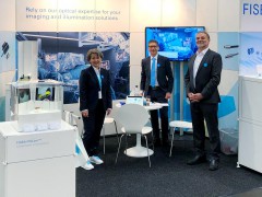 FISBA exhibits Customized Solutions for Minimally Invasive Medical Endoscopic Devices at COMPAMED in
 New Active Alignment System for the Coupling of Photonic Structures to Fiber Arrays
 A new industrial compression module by Amplitude
 Menhir Photonics Introduces the MENHIR-1550 The Industry's First Turnkey Femtosecond Laser of
 Shenzhen DNE Laser introduced new generation D-FAST cutting machine (12000 W)