Kelheim, Germany, 31. August 2015.
With the launch of its pco.flim system, PCO AG in Bavaria, Germany, is ushering in a new era of fluorescence microscopy. The camera has a revolutionary image sensor and makes use of fluorescence lifetime imaging in the frequency domain, making it suitable for numerous applications in the field of biomedical research. Dr Gerhard Holst, Head of Research at PCO, explains more: ?The pco.flim system is the result of great tenacity and the support of research projects over the last eight years as we have worked towards series production. This development yet again highlights PCO?s position as one of the global leaders for the production of scientific cameras.?
The secret of this camera?s success is the charge-swing in its sensor. In normal camera chips, the light creates charge carriers that are recorded in a detection bin? one per pixel. The quantity of charge carriers and thus the brightness of each pixel is measured electronically. The pco.flim system has two of these bins per pixel, which can be switched at an incredible speed. This makes it possible to do more than simply measure the total quantity of light ? it is also possible to sample light information over time.
This is of particular significance in fluorescence microscopy, which is widely used in the fields of cell research and molecular medicine. An example of its use is when cancer cells are marked with a fluorescent dye. The dye is luminous when it is exposed to excitation light coming for example from a laser diode. The brightness distribution provides information about the cell?s structure and metabolic processes. Most fluorescence microscopes are equipped with cameras that simply measure light distribution. Fluorescence lifetime imaging microscopy (or FLIM for short) is much rarer and more advanced. It involves modulating the excitation light, which means that the dye in the cell then also returns modulating light, with a slight time delay.. This time delay provides further useful information about the metabolic processes in the cell. Until now, cameras that are also capable of sampling the light signal over time have been too slow and have required additional equipment. Due to limited results and high costs, FLIM has remained a niche application.
With the arrival of the pco.flim system, this will no longer be the case. It is the first camera that combines all of the relevant requirements. Its resolution of 1008 x 1008 pixels means that the images it takes are sharp. The pixels can be switched at a frequency of up to 40 MHz and this can be synchronised with the modulation of the excitation light. This means that the camera can even detect delays of less than 100 picoseconds (100 trillionths of a second) between the excitation light and the fluorescence. Another advantage is that the pco.flim system only requires minimal additional equipment. All this means that a FLIM microscope with a PCO camera will be less than half the price of existing systems, while offering better performance.
The Kelheim-based firm will officially present the pco.flim system to the public at the 14th Conference on Methods and Applications of Fluorescence, which is taking place in W?zburg, Germany from 13-16 September 2015.
An overview of key parameters for the PCO FLIM:
- Resolution of 1008 x 1008 pixels.
- 90 double images per second.
- Modulation frequency range of 5 kHz?40 MHz.
- Sine and square wave modulation forms.
- Readout noise of 13 electrons (med).
- Dynamic range of > 1000:1.
- Quantum efficiency of 39%.
- Rolling shutter readout mode.

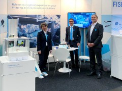 FISBA exhibits Customized Solutions for Minimally Invasive Medical Endoscopic Devices at COMPAMED in
 New Active Alignment System for the Coupling of Photonic Structures to Fiber Arrays
 A new industrial compression module by Amplitude
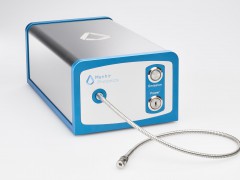 Menhir Photonics Introduces the MENHIR-1550 The Industry's First Turnkey Femtosecond Laser of
 Shenzhen DNE Laser introduced new generation D-FAST cutting machine (12000 W)